Last weeks description of the challenges in using neuro data in education.
Day 1: Basic Architecture
Day 2: Single Cell Inspiration
Most of the time, neuroscientific research is designed to tell us something about the brain, or about brain-behavior relationships. Occasionally, we are in a position to use neuroscientific data to inform purely behavioral theories.
A great example of this principle comes from the debates of the 1970's and 1980's regarding the basis of visual mental imagery.

During the 1970's there was a lively (often acrimonious) debate as to the mental representation that allows people to answer this sort of question. Some researchers (most visibly, Stephen Kosslyn) argued that we use mental representations that are inherently spatial, and that visual mental imagery overlaps considerably with visual perception.
Other researchers (most visibly, Zenon Pylyshyn) argued that the feeling of inspecting an image may be real, but the inspection does not support your answering the question. The mental representation that allows you to say "the torch is in her right hand" is actually linguistic , and therefore has a lot in common with the language system.
Although these two theories seem radically different, it was actually quite difficult (technically it was impossible--Anderson, 1978) to distinguish between them via behavioral experiments only.
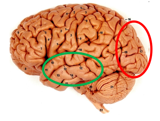
But even 30 years ago, we knew enough about the brain to gather neuroscientific data that helped to decide between these competing theories. We knew some of the key brain areas supporting visual perception (red circle below) and we knew some of those supporting language representations (green circle).
Functional brain imaging makes corresponding predictions about which parts of the brain will be active during imagery tasks, and again, the data supported the theory positing that imagery relies on representations similar to those supporting visual perception (e.g., Le Bihan et al, 1993).
Can this technique be applied to education? Yes.
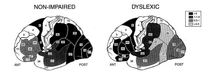
For example, Shaywitz et al (1998) imaged the brains of typical readers and readers diagnosed with dyslexia during tasks of increasing phonological complexity. A number of differences in activation emerged, as shown below (click for larger image).
What's especially interesting from our perspective today is that the researchers interpreted these data in light of other data leading them to suggest that they already knew what certain brain areas do.
Thus, the authors highlighted the abnormal activity in area 39 (the angular gyrus) which they suggested is crucial for translating visual input into sound; they pointed to other research showing that people who were typical readers suddenly had trouble reading if that brain area suffered damage.
Thus another way we can use neuroscientific data is to leverage what we already know about the brain to test behavioral theories.
We have to not that it's also pretty easy to get fooled with this method. Obviously, if we think we know what a brain area supports but we're wrong, we'll draw the wrong conclusion. Then too, we might be right about what the brain area supports, but it may support more than one behavior.
For example, there are good data showing that the amygdala is important in the processing of some emotions, especially fear. But it would be hasty (and wrong) to suppose that every time the amygdala is active, it's because the person is experiencing strong emotion. Amygdala activation is observed in many circumstances, and it very likely participates in a number of functions.
So using our knowledge of the brain to inform behavioral theories is great, but it's easy to screw up.
Tomorrow: the most common misconception about brain imaging and how to correct it.
Anderson, J. R. (1978). Arguments concerning representation for mental imagery. Psychological Review, 85, 249-277.
Farah, M. J. (1988). Is visual mental imagery really visual? Overlooked evidence from neuropsychology. Psychological Review, 95, 307-317.
Le Bihan, D, Turner, R., Zeffiro, T. A., Cuenod, C. A., Jezzard, P. & Bonnerot, V. (1993) Activation of human primary visual cortex during visual recall: A magnetic resonance imaging study. Proceedings of the National Academy of Sciences, 90, 11802-11805.
Shaywitz, S. E et al (1998). Functional disruption in the organization of the brain for reading in dyslexia. Proceedings of the National Academy of Sciences, 95, 2636-2641.


 RSS Feed